
Canine otitis externa (OE) remains one of the most frequent presentations in veterinary practice, accounting for up to 15–20% of canine consultations [1]. Historically, OE was often approached empirically—treated as a simple superficial infection with broad-spectrum “shotgun” topical therapies. However, rising antimicrobial resistance (AMR), an enhanced understanding of otic microbiology, and advancements in drug delivery systems have reshaped the clinical standard of care.
Today, successfully managing canine otitis externa requires moving beyond managing symptoms. It demands a systematic, multi-factorial approach, strict antimicrobial stewardship, and aggressive anti-inflammatory management to prevent progression to irreversible end-stage disease.
1. The Diagnostic Foundation: The PSPP System
Treating the secondary infection without resolving the underlying cause is one of the most common reasons for treatment failure and recurrence. Modern veterinary otology categorizes every case through the PSPP System [2, 3]:

Fig 1: Gemini AI.(2006, August 10). The PSPP Diagnostoc System for Canine Otits Externa[AI-generated image]. Created using Gemini image generation tool.
Primary Causes: These initiate the inflammatory cascade in a previously healthy ear canal.
- Hypersensitivity Disorders: Canine Atopic Dermatitis (CAD) and cutaneous Adverse Food Reactions (cAFR) account for up to 75–80% of recurrent cases. In fact, up to 24% of dogs with AFR present with otitis externa as their sole clinical complaint [4].
- Foreign Bodies & Parasites: Grass awns, and ear mites (more common in young dogs).
- Endocrinopathies: Hypothyroidism and hyperadrenocorticism alter epidermal turnover and cerumen lipid composition, predisposing the canal to microbial proliferation.
Predisposing Factors: These do not cause otitis on their own, but increase susceptibility.
- Anatomy: Pendulous pinnae (e.g., Basset Hounds), narrow canals (e.g., Shar-Pei’s), and excessive intraluminal hair (e.g., Poodles) restrict airflow and trap moisture.
- Micro-environment: Swimming, frequent bathing, or humid environments cause maceration of the stratum corneum.
- Genetic predisposition: Some breeds like the American Cocker Spaniel are genetically more prone to developing allergic OE.
Secondary Causes: These are opportunistic pathogens that proliferate in damaged otic micro-environments.
- Yeasts: Malassezia pachydermatis.
- Bacteria: Gram-positive cocci (Staphylococcus pseudintermedius) and Gram-negative rods (Pseudomonas aeruginosa, Proteus spp, Corynebacterium).
Perpetuating Factors: These develop secondary to chronic inflammation and prevent resolution.
- Pathological Changes: Epithelial hyperplasia, dermal oedema, ceruminous gland hypertrophy, luminal stenosis, and dystrophic follicular mineralization.
- Otitis Media (OM): Concurrent middle ear disease is present in up to 16% of acute cases and over 50% of chronic/recurrent cases [5, 6]. The bulla acts as a persistent reservoir for reinfection if the tympanic membrane is ruptured or incompetent.
2. Modern Diagnostic Workflow
A structured diagnostic protocol prevents diagnostic oversights and guides targeted therapy:
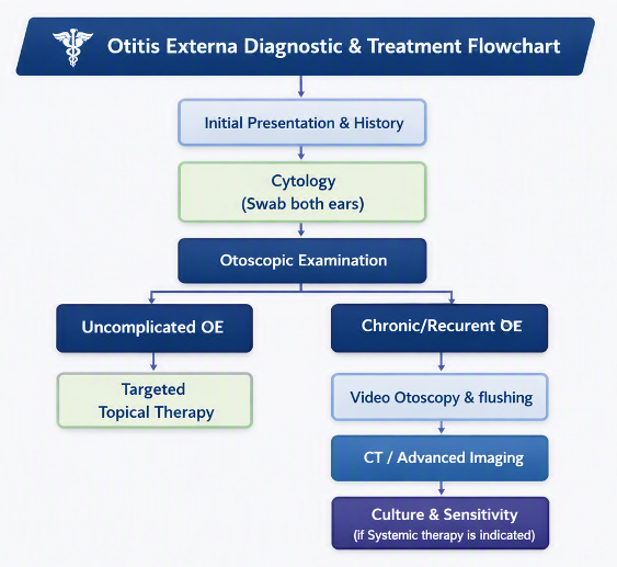
Fig. 2: Microsoft Copilot.(2026, August 11). Otits Externa Diagnostic & Treatment Flow chart [AI-generated image]. Created using Microsoft Copilot graphic art tool.
In-Clinic Otic Cytology: Non-Negotiable at Every Visit
Visual inspection alone cannot distinguish between bacterial and yeast infections or predict the nature of the cellular response. Quantitative cytology must be performed at initial diagnosis and at every re-check examination until cytological resolution is achieved [1].
- Sample Collection: Roll (do not smear) cotton swabs onto a clean glass slide, heat-fix lightly if high wax content is present, and stain using a modified Wright-Giemsa stain (e.g., Diff-Quik).
- Quantification: Evaluate under high-power oil immersion.
- Malassezia: Peanut-shaped budding yeasts (>2–5 per high-power field [HPF] is typically considered abnormal depending on the breed).
- Cocci & Rods: Note presence, relative abundance, and intracellular versus extracellular locations.
- Inflammatory Cells: Presence of degenerate neutrophils indicates active tissue infection rather than simple overgrowth. Phagocytosed bacteria confirm an active infection.
The Role and Nuance of Culture & Susceptibility (C&S)
Bacterial culture is often over-utilized or misapplied in OE management [7].
Key Clinical Concept: Standard laboratory susceptibility breakpoints are based on achievable systemic (plasma) drug concentrations. Topical medications deliver drug concentrations to the ear canal that are 100 to 1,000 times higher than systemic MIC thresholds [8]. Therefore, an organism reported as “resistant” in vitro may still be eliminated by topical therapy.
When is C&S Indicated?
- When systemic antimicrobial therapy is indicated (e.g., concurrent otitis media, marked soft tissue swelling/ulceration of the canal).
- Persistent rod-shaped bacteria despite appropriate empirical topical therapy.
- Cytological evidence of persistent infection alongside severe, erosive, or ulcerative otitis.
Video Otoscopy and Middle Ear Evaluation
A handheld otoscope offers a limited field of view in stenotic or debris-filled canals. High-definition video otoscopy (VO) has transformed veterinary otology [6]:
- Allows precise visualization of the deep vertical/horizontal canals and the tympanic membrane (TM).
- Facilitates deep ear flushing under general anesthesia to remove tenacious ceruminous or purulent plugs.
- Enables myringotomy and middle ear aspiration/flushing when otitis media is confirmed or suspected.
In cases where middle ear pathology, tissue proliferation, or bony changes are suspected, computed tomography (CT) is significantly more sensitive than radiography for evaluating bulla wall thickening, soft tissue attenuation, and osteolysis [9].
3. Novel & Targeted Therapeutic Strategies
Antimicrobial Stewardship and Antibiotic-Free Formulations
The widespread use of combination products containing an antibiotic, an antifungal, and a corticosteroid for every ear complaint has contributed to rising multidrug-resistant pathogens. However, in a recent study, 38.9% of canine OE cases did represent a mixed infection (M. intermedius and Staphylococci spp) [13] and all cases where skin lesions were present showed M. pachydermitis isolated together with a baterial pathogen[13]. Recent advances support a more evidence-based approach:
- Targeted Antifungal Therapy: For ears demonstrating pure Malassezia overgrowth on cytology without bacterial involvement, using broad-spectrum antibiotic combinations is unnecessary. The availability of antibiotic-free, targeted formulations—such as FDA-approved terbinafine and betamethasone acetate gel (DuOtic)—delivers potent antifungal and anti-inflammatory action while avoiding unnecessary antibiotic exposure.
- Mono-Infection Target Selection: Select topical drops containing single antimicrobial agents matched directly to cytology rather than default triple-combination products when possible.
Long-Acting In-Clinic Delivery Systems
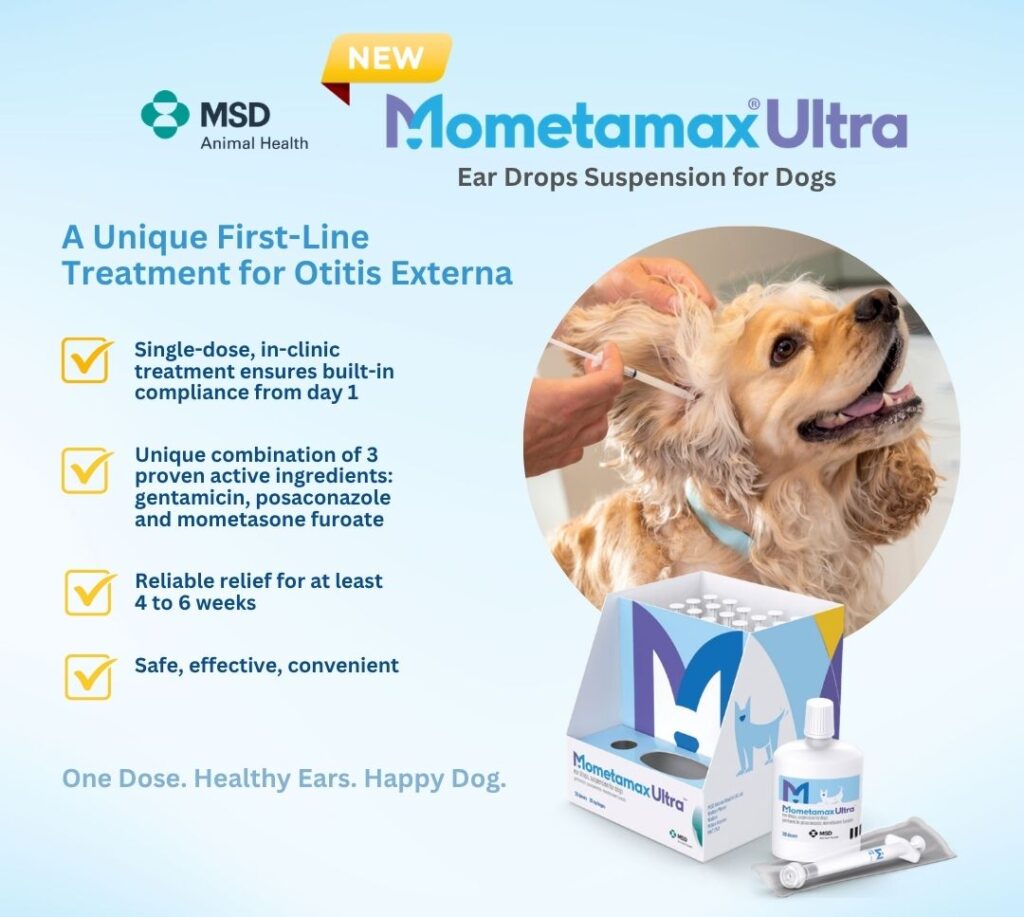
Client compliance failure (under-dosing, stopping therapy early due to pain, or incorrect administration technique) represents a leading driver of therapeutic failure [1]. Long-acting solutions (e.g., gentamicin/posaconazole/mometazone furoate combination administered directly by veterinary staff can reshape compliance dynamics and improve resolution outomes.
- Mechanism: These bioadhesive formulations cross-link or adapt to ear wax and exudate, sustainably releasing active ingredients over extended periods.
- Protocol: Initial in-clinic ear cleaning and removal of any bacterial biofilm along with drying prior to application. Ears should not be flushed or cleaned at home during the treatment window to avoid washing out the sustained-release matrix.
Managing Biofilms and Refractory Pseudomonas
Pseudomonas aeruginosa is a formidable pathogen in veterinary otology due to intrinsic efflux pumps, low membrane permeability, and its propensity to produce extracellular polymeric substances (biofilms) [10]. Biofilms act as physical barriers, shielding bacteria from both topical antimicrobials and host immune defenses.
Biofilm Disruption Protocol:
- Tris-EDTA (Tromethamine-EDTA): Tris-EDTA chelates calcium and magnesium ions in bacterial cell walls and biofilm matrices, dramatically increasing cell membrane permeability [11]. Administer Tris-EDTA 15 – 30 minutes before applying topical antibiotics (such as fluoroquinolones or aminoglycosides) to produce a synergistic bactericidal effect.
- N-Acetylcysteine (NAC) & Mucolytics: 2% NAC solutions help break down mucopurulent exudate and biofilm architecture during deep canal flushing.
- Topical Fluoroquinolones & Aminoglycosides: High-concentration topical gentamicin and marbofloxacin remain frontline choices once the biofilm is disrupted.
4. Anti-Inflammatory Management: Reversing Pathological Changes
Antimicrobials address secondary microbial overgrowth, but anti-inflammatory therapy represents a key pilllar in controlling the core pathology of otitis externa. Uncontrolled inflammation initiates a cycle of oedema, hyperplasia, stenosis, and eventually, irreversible canal mineralization.
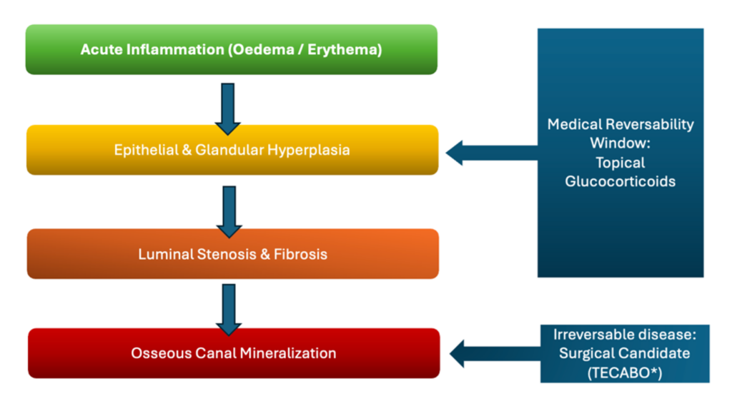
Fig. 3: M.Enslin. (2026, August 17). Inflammation Progression Diagram (Otitis Externa) [Image created in Microsoft PowerPoint]. Based on Rosychuk, R.A.W. (1994). Otitis Externa: Diagnosis and Medical Management. Veterinary Clinics of North America: Small Animal Practice, 24(5), 921-952.
*Total Ear Canal Ablation and Bulla Osteotomy.
Glucocorticoid Protocols
- Topical Glucocorticoids: Hydrocortisone or prednisolone may suffice for mild, acute inflammation. However, moderate-to-severe stenotic canals require high-potency topical agents such as mometasone furoate, betamethasone, or dexamethasone.
- Systemic Glucocorticoids: When marked luminal stenosis prevents topical drops from penetrating the horizontal canal, a short course of oral prednisone or prednisolone (0.5–1.0 mg/kg q24h for 5–10 days) is indicated [3]. Reopening the canal restores ventilation and allows topical medications to reach the deep horizontal canal and tympanic membrane.
Recognizing the Point of No Return: Mineralization vs. Surgery
Once chronic inflammation leads to complete dystrophic calcification and mineralization of the auricular cartilage, the canal loses all physiologic function.
- Palpation: Healthy canal cartilage is flexible and pliable. Mineralized canals feel rigid, unyielding, and bone-like on external palpation [1].
- Management Shift: Ears with end-stage mineralized canals cannot be managed medically. Attempting medical therapy in these ears causes chronic pain and invites recurrent resistant infections. Total Ear Canal Ablation with Lateral Bulla Osteotomy (TECABO) is the humane, definitive surgical recommendation to relieve chronic pain [12].
Summary of Clinical Best Practices
| Clinical Pillar | Evidence-Based Recommendation | Key Pitfall to Avoid | References |
|---|---|---|---|
| Diagnostics | Perform cytology at initial evaluation and every recheck until cytological resolution. | Discontinuing treatment based solely on visual improvement. | [1, 3] |
| Culture Testing | Reserve C&S for systemic therapy cases, persistent rods, or deep tissue ulceration. | Assuming topicals are ineffective due to in vitro systemic MIC resistance. | [7, 8] |
| Biofilm Control | Pre-treat purulent or Pseudomonas ears with Tris-EDTA 15–30 minutes prior to antibiotics. | Applying topical antimicrobials directly over thick debris or biofilm. | [10, 11] |
| Long-Term Control | Identify and manage primary drivers (atopy, AFR, hypothyroidism) alongside infection. | Treating recurring infections as “isolated events” without investigating underlying causes. | [2, 4] |
References
- Nuttall, T. (2016). Successful management of otitis externa in dogs. In Practice, 38(Suppl 2), 17–22.
- Paterson, S., & Matousek, J. L. (2013). Otitis Externa in the Dog and Cat. Royal Society of Medicine Press.
- Bajwa, J. (2019). Canine otitis externa – Treatment and complications. Canadian Veterinary Journal, 60(1), 97–99.
- Favrot, C., Steffan, J., Seewald, W., & Picco, F. (2010). A prospective study on the clinical features of chronic canine atopic dermatitis. Veterinary Dermatology, 21(1), 23–31.
- Cole, L. K. (2012). Anatomy and physiology of the canine ear. Veterinary Dermatology, 23(1), 3–12.
- Gotthelf, L. N. (2004). Diagnosis and treatment of otitis media in dogs and cats. Veterinary Clinics of North America: Small Animal Practice, 34(2), 469–487.
- Morris, D. O. (2019). Medical management of canine otitis externa and otitis media. Veterinary Clinics of North America: Small Animal Practice, 49(5), 761–774.
- Frank, L. A., Kania, S. A., Hnilica, K. A., & Wilkes, R. P. (2003). Isolation of Pseudomonas aeruginosa from dogs with otitis externa and susceptibility to topical antimicrobial agents. Journal of the American Veterinary Medical Association, 223(7), 1000–1002.
- Belmudes, A., Pressanti, C., Barthez, P. Y., Cauzinille, L., & Bouvy, P. (2018). Computed tomographic findings in 205 dogs with otitis media. Veterinary Radiology & Ultrasound, 59(1), 32–42.
- Pye, C. C. (2018). Pseudomonas otitis externa in dogs. Canadian Veterinary Journal, 59(11), 1231–1234.
- Buckley, C. L., Schmidt, V. M., McEwan, N. A., & Nuttall, T. (2013). Cross-sectional study of resistance to antimicrobials in bacterial isolates from dogs with otitis externa or otitis media. Veterinary Record, 172(12), 312.
- Mason, C. L., Simpson, D. M., & Hall, J. L. (2013). Long-term outcome following total ear canal ablation and lateral bulla osteotomy in dogs. Journal of Small Animal Practice, 54(4), 193–199.
- Dinkova V, Rusenova N. Prevalence of Malassezia pachydermatis yeasts in clinical infections in dogs: A single-center retrospective study at the University Veterinary Hospital, Stara Zagora, Bulgaria (2019-2023). Open Vet J. 2025 Sep;15(9):4354-4361. doi: 10.5455/OVJ.2025.v15.i9.42. Epub 2025 Sep 30. PMID: 41200355; PMCID: PMC12587832.
NB: Products mentioned or referred to in the blog:
1: Mometemax Ultra
2: Tris-Nac (part of the ICF range)